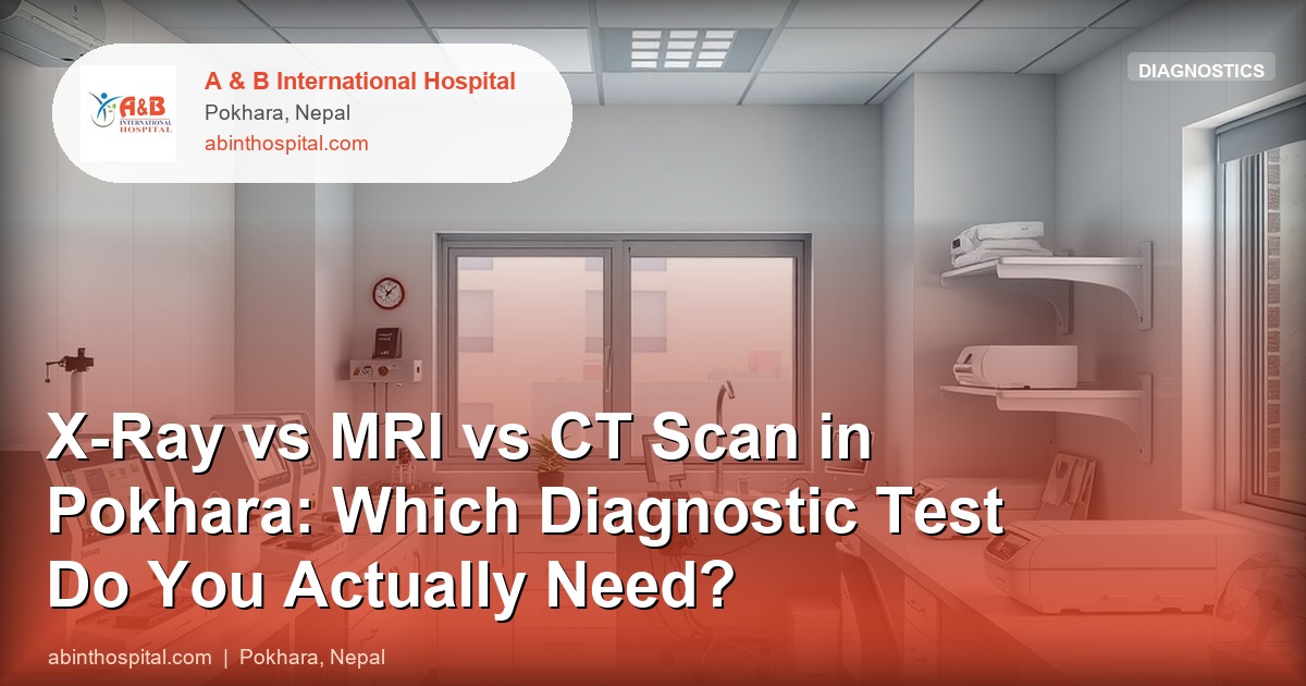
X-Ray vs MRI vs CT Scan in Pokhara: Which Diagnostic Test Do You Actually Need?
Patients in Pokhara are frequently referred for imaging but are often unsure which test does what, which carries radiation, and which is available locally versus requiring referral. This guide provides a factual comparison of digital X-ray, CT scan, and MRI — what each modality shows, when it is the right choice, and how to make sense of your imaging report.
What Is Digital X-Ray and What Does It Show Best?
X-ray uses ionising radiation transmitted through the body. Dense structures such as bone absorb the radiation and appear white on the image. Air-filled structures such as the lungs appear black. Soft tissues fall in between as shades of grey.
Digital X-ray at A&B provides immediate, high-resolution images for:
- Bone injuries: Fractures, dislocations, joint abnormalities, bone tumours
- Chest pathology: Pneumonia (lung consolidation), pleural effusion (fluid around the lung), pneumothorax (collapsed lung), cardiomegaly (enlarged heart), pulmonary tuberculosis
- Abdominal emergencies: Perforation (free air under diaphragm), bowel obstruction
- Spine: Degenerative changes, scoliosis, compression fractures
X-ray is the appropriate first-line imaging test for suspected fractures, chest infections, and pre-operative workup. The radiation dose from a chest X-ray is approximately 0.1 mSv — equivalent to about 10 days of natural background radiation. A limb X-ray delivers even less.
Digital radiography reduces patient radiation exposure by 50–80% compared to conventional film-screen X-ray because image quality is digitally adjustable and repeat exposures for positioning errors are rare.
What Does a CT Scan Show?
CT (Computed Tomography) uses multiple X-ray beams rotating around the patient to generate detailed cross-sectional images of any body part. It shows both bone and soft tissue in the same scan, with excellent spatial resolution. CT is significantly faster than MRI — a head CT takes approximately 30 seconds, making it ideal for emergencies.
CT is best for:
- Emergency brain assessment: Ischaemic stroke vs haemorrhagic stroke (distinguishing the two is essential before starting blood thinners), head injury (subdural haematoma, extradural haematoma, cerebral contusion)
- Chest CT: Pulmonary embolism (CT pulmonary angiography), lung tumours, complex chest infections, aortic aneurysm
- Abdominal CT: Appendicitis, kidney stones, abdominal aortic aneurysm, bowel tumours, liver masses
- Trauma: Whole-body trauma CT for major accidents
- Bone detail: Complex fractures (especially spine, pelvis, wrist), tumour invasion
Radiation dose: A head CT delivers approximately 2 mSv; an abdominal CT delivers 5–10 mSv. CT should not be ordered unless clinically necessary — but when it is needed (particularly in emergencies), its speed and detail are essential.
Contrast agents: Iodinated contrast dye is often injected intravenously to enhance blood vessels and detect tumours or infections. Before contrast administration, renal function (creatinine) should be checked, as iodinated contrast can cause kidney injury in patients with renal impairment. Patients with contrast allergy or borderline renal function need pre-medication or alternative strategies.
CT scanning is not available at A&B. Patients requiring CT are referred to appropriate CT-equipped facilities in Pokhara. A&B doctors provide clinical assessment, initiate basic stabilisation, and arrange appropriate referral with all prior investigation results.
What Does an MRI Show?
MRI (Magnetic Resonance Imaging) uses strong magnetic fields and radio waves — no ionising radiation. It produces exquisitely detailed soft tissue images, making it superior to CT for most brain, spinal cord, joint, and soft tissue assessments.
MRI is best for:
- Brain: Tumours, multiple sclerosis, posterior fossa lesions (which CT shows poorly), encephalitis, early ischaemic stroke after the first 24–48 hours
- Spine: Intervertebral disc prolapse (the definitive test for back pain with radiculopathy), spinal cord compression, infection of vertebrae (discitis/osteomyelitis)
- Joints: Meniscal tears, anterior cruciate ligament (ACL) tears, rotator cuff tears — A&B’s physiotherapy team uses MRI reports to plan rehabilitation
- Soft tissue: Muscle tears, abscesses, soft tissue tumours
- Pelvic organs: Uterine and cervical pathology where detail beyond ultrasound is needed
Radiation dose: Zero — MRI uses no radiation. It can be repeated without radiation concern.
Contraindications:
- Absolute: Pacemakers (older non-MRI-compatible devices), cochlear implants, some older aneurysm clips, metallic intraocular foreign bodies. These are strict contraindications — MRI cannot be performed.
- Relative: Other metallic implants (most modern orthopaedic implants are MRI-safe, but must be checked), severe claustrophobia.
MRI is not available at A&B but is accessible in Pokhara through referral. A&B coordinates referrals with imaging centres in the city.
Cost Comparison in Pokhara (Approximate)
| Modality | Approximate Cost Range |
|---|---|
| Digital X-ray (single view) | Rs 300–800 |
| Ultrasound (abdomen) | Rs 1,500–3,000 |
| ECG | Rs 400–800 |
| CT scan (head, non-contrast) | Rs 5,000–10,000 |
| CT scan (abdomen with contrast) | Rs 10,000–18,000 |
| MRI (brain) | Rs 12,000–20,000 |
| MRI (lumbar spine) | Rs 12,000–18,000 |
Costs vary by institution and indication. ECHS covers approved imaging investigations at empanelled rates for entitled beneficiaries.
Which Modality Should You Use for Common Clinical Situations?
| Clinical Question | Recommended First Test |
|---|---|
| Suspected fracture (limb, rib) | X-ray |
| Chest infection, TB | Chest X-ray |
| Suspected stroke (emergency) | CT head immediately |
| Head injury with loss of consciousness | CT head immediately |
| Back pain with leg weakness or numbness | MRI lumbar spine |
| Kidney stones | Ultrasound first; CT KUB if inconclusive |
| Appendicitis (clinical diagnosis uncertain) | CT abdomen |
| Knee pain with suspected ligament/meniscal tear | MRI knee |
| Abdominal pain with suspected gallstones | Ultrasound abdomen |
| Pulmonary embolism suspicion | CT pulmonary angiography |
| Palpable lump (soft tissue) | Ultrasound first; MRI for characterisation |
The overarching principle is appropriate imaging — not the most expensive test, but the right test for the right question. Using MRI for every back pain case is wasteful and not more informative than X-ray if the clinical question is simply whether a fracture is present.
What Imaging Is Available Directly at A&B?
A&B International Hospital provides digital X-ray and ultrasound on-site with same-day results. Echocardiography (cardiac ultrasound) is also available. For CT and MRI, A&B clinicians assess the patient, conduct available diagnostics, stabilise if needed, and coordinate prompt referral to Pokhara’s CT/MRI facilities with all relevant clinical notes.
Book Imaging at A&B International Hospital
A&B International Hospital
Pokhara-02, Bindhyaabasini Way to Sarangkot
Phone: +977 061-412512
Digital X-ray and ultrasound with same-day results. CT and MRI referral coordination available. ECHS imaging services for entitled beneficiaries. 24/7 emergency imaging access.