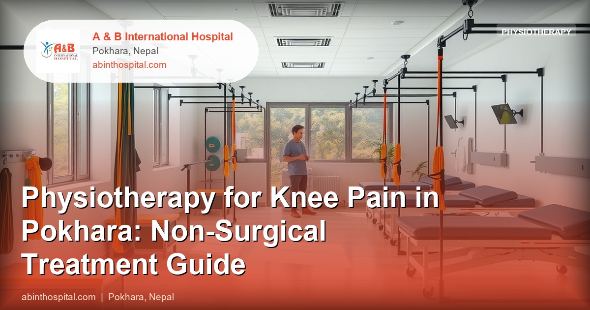
Physiotherapy for Knee Pain in Pokhara: Non-Surgical Treatment Guide
Knee pain is among the most common musculoskeletal complaints in the Pokhara region, affecting agricultural workers who spend hours in squatting postures, older adults with osteoarthritis, veterans with occupational injury histories, and younger patients with sports and trekking injuries. The knee is a complex joint that is poorly served by generic pain management — different knee conditions require fundamentally different physiotherapy approaches.
What Knee Conditions Can Physiotherapy Treat?
Knee osteoarthritis (OA) is the most prevalent knee condition in adults over 50 in Nepal. It involves progressive cartilage loss in the joint, causing pain, stiffness, and loss of function. Physiotherapy is the cornerstone of evidence-based management for knee OA and is recommended as first-line treatment by NICE, EULAR, and Osteoarthritis Research Society International (OARSI) guidelines — before considering surgery.
Patellofemoral pain syndrome (PFPS) — pain around or behind the kneecap — is common in younger adults and runners. It is caused by abnormal kneecap tracking related to hip and quad weakness, tight lateral structures, and poor neuromuscular control. It responds very well to targeted physiotherapy.
Meniscus irritation from degenerative tears (common in older adults with OA) or acute tears (in younger athletes following twisting injury) causes pain at the joint line, swelling, and sometimes mechanical locking. Physiotherapy is effective for degenerative meniscus tears; acute traumatic tears in young athletes often require surgical assessment.
IT band syndrome produces lateral knee pain in runners and cyclists. The iliotibial band — a thick band of fascia running from the hip to the outer knee — becomes irritated where it crosses the lateral femoral condyle. Treatment focuses on hip abductor strengthening and running biomechanics.
Patellar tendinopathy (jumper’s knee) is characterised by inferior patellar pain with loading activities — jumping, squatting, stair climbing. It is common in volleyball, basketball, and heavy trekking. It responds specifically to heavy slow resistance (HSR) exercise protocol applied to the tendon.
How Is Knee Pain Assessed at A&B Physiotherapy?
Assessment begins with determining which structure is causing the pain. The location of pain provides the first clue: anterior (patellofemoral, patellar tendon), medial (medial compartment OA, medial collateral ligament, medial meniscus), lateral (IT band, lateral compartment OA, lateral meniscus), or posterior (Baker’s cyst, popliteus tendon).
Special tests clarify: the Valgus stress test for MCL integrity, McMurray’s test for meniscus, Patellar grind/Clarke’s test for patellofemoral pain, Lachman’s test for ACL integrity. Movement assessment identifies muscle imbalances: hip abductor weakness causing dynamic valgus (knee caving inward), quadriceps weakness causing increased knee loading, and calf tightness increasing patellar tracking problems.
Understanding the mechanism — how the knee came to be painful — is equally important. A sudden painful knee after twisting in a sports match suggests ligament or meniscus injury. Gradual onset bilateral knee pain after age 50 in an overweight patient with morning stiffness suggests osteoarthritis.
What Physiotherapy Techniques Are Used for Knee Pain?
Quadriceps strengthening is the most important intervention for virtually all knee conditions. The quadriceps are the primary shock absorbers of the knee — every 1 kg of quadriceps strength reduces the load on the knee joint by 3–4 kg. Weakness is nearly universal in patients with knee pain from any cause, partly from pain inhibition and partly from deconditioning.
Vastus medialis oblique (VMO) activation is specifically targeted in patellofemoral pain — this medial portion of the quadriceps pulls the patella medially and is often preferentially weakened, causing the patella to track too far laterally. Terminal knee extension exercises and mini-squats with emphasis on last-range extension specifically train the VMO.
Manual therapy — patellar mobilisation, tibiofemoral joint mobilisation, soft tissue work to the IT band and lateral structures — improves joint mechanics and reduces pain acutely. It is most effective as a preparation for exercise.
Patellar taping using McConnell technique temporarily corrects patellar tracking, providing pain relief that allows the patient to exercise more effectively. It is not a standalone treatment but a useful adjunct.
Hip strengthening — specifically gluteus medius (hip abductor) and gluteus maximus — reduces dynamic valgus collapse during weight-bearing, decreasing medial compartment load and patellofemoral stress. Hip strengthening is a component of treatment for nearly all knee conditions.
What Is the Impact of Weight on Knee Pain?
Body weight has a disproportionate mechanical effect on the knee. The knee joint carries 3–6 times body weight during walking and up to 8 times during stair climbing. A 5 kg weight reduction produces a 15–30 kg reduction in knee joint load with every step.
This is not a judgment about the patient — it is a mechanical reality that physiotherapists discuss directly with patients who are overweight or obese. Weight management is a joint-protection strategy, not a cosmetic aspiration. Combined physiotherapy and weight management produces better outcomes for knee OA than either alone.
When Is Physiotherapy Not Enough and Surgery Is Needed?
For knee OA, surgery — total or partial knee replacement — is indicated when pain is severe, function is substantially impaired, quality of life is significantly affected, and adequate non-surgical management (physiotherapy, weight management, anti-inflammatory medication, intra-articular injections) has been tried and has not provided sufficient relief.
For meniscus tears in young patients with mechanical symptoms (locking, giving way, acute swelling), arthroscopic surgery may be appropriate. For ACL tears in patients returning to pivoting sport, surgical reconstruction is generally recommended.
Physiotherapy both before and after knee surgery improves outcomes. Pre-operative physiotherapy (“prehabilitation”) — building quadriceps and hamstring strength before a planned knee replacement — improves post-operative function and speeds recovery.
What Is the Exercise Progression for Knee Osteoarthritis?
Week 1–2: Seated knee extensions (bodyweight), straight leg raises, static quad sets, heel slides for flexion range, gentle cycling on stationary bike if available.
Week 3–4: Mini squats to 30 degrees (pain-free range), step-ups on a low step, calf raises, short walks on level ground.
Week 5–8: Squat depth progressively increased, longer walks, gentle stair work, resistance band leg press.
Months 3+: Functional strengthening, progressive resistance, return to previous activities progressively.
All exercises are prescribed and monitored by the physiotherapist, with progression guided by pain response (no more than 3–4/10 pain during exercise, returning to baseline within 24 hours).
A&B International Hospital
Pokhara-02, Bindhyaabasini Way to Sarangkot
Phone: +977 061-412512
Website: abinthospital.com
Knee pain does not necessarily mean knee surgery. A physiotherapy assessment at A&B International Hospital, Pokhara will identify the cause of your knee pain and develop a structured, evidence-based treatment plan. ECHS polycards accepted for eligible veterans.